
Description
EHD refers to Hemorrhagic Disease, an illness that causes extensive bleeding and is sometimes fatal in deer. It is caused by two groups of related viruses: Epizootic Hemorrhagic Disease (EHD) viruses and Bluetongue Viruses (BT). Hemorrhagic Disease in Michigan white-tailed deer is almost always caused by the EHD group of viruses, which is why the name is often shortened to “EHD”. Over the past two decades, this condition has been responsible for substantial outbreaks in deer in many northern U.S. states and provinces in southern Canada. Throughout the U.S., most animals affected by EHD are white-tailed deer, but outbreaks in mule deer and pronghorn have also occurred.
Distribution
Although EHD was likely present in North America long before it was recognized, it was first identified in 1955 when several hundred white-tailed deer (Odocoileus virginianus) succumbed in New Jersey and Michigan. At the time, it was considered a new disease of deer, and the name 'epizootic hemorrhagic disease' was given to the condition.
Over the next several decades, EHD was identified in multiple locations in North America, but only one other outbreak was detected in Michigan. It recurred in Michigan in 2006 and has been identified here nearly every year since.
Since first diagnosed, mortality events attributed to EHD occurred in Michigan in white-tailed deer in the following counties:
• 1955 — Lake, Manistee, Muskegon, Saginaw
• 1974 — Arenac, Ingham, Iosco, Gratiot and Mecosta.
• 2006 — Allegan.
• 2008 — Oakland and Macomb.
• 2009 — Livingston.
• 2010 — Allegan, Berrien, Cass, Ottawa, St. Joseph and Van Buren.
• 2012 — Allegan, Barry, Branch, Calhoun, Cass, Clare, Clinton, Eaton, Genessee, Gratiot, Hillsdale, Ingham, Ionia, Isabella, Jackson, Kalamazoo, Kent, Lenawee, Manistee, Mecosta, Montcalm, Muskegon, Newago, Osceola, Ottawa, Saginaw, Shiawassee, St. Joseph, Washtenaw and Wayne.
• 2013 — Allegan, Berrien, Ionia, Muskegon, Oceana, Ottawa and St. Joseph.
• 2014 and 2015 — no cases reported.
• 2016 — Berrien, Cass and Van Buren.
• 2017 — Allegan, Barry, Calhoun, Genesee, Hillsdale, Jackson, Lenawee, Livingston, Montcalm, Saginaw, Shiawassee, St. Clair and Washtenaw.
• 2018 — Calhoun and Kent.
• 2019 — Cass.
• 2020 — Allegan, Kalamazoo, Roscommon and Saginaw.
• 2021 — Berrien, Branch, Macomb, Oakland, Shiawassee and St. Clair.
• 2022 — Cass, Lenawee, Montmorency, Ogemaw and Washtenaw.
• 2023 — Washtenaw.
• 2024 — Allegan, Barry, Berrien, Branch, Calhoun, Cass, Genesee, Hillsdale, Jackson, Kalamazoo, Kent, Lenawee, Ottawa, St. Joseph and Van Buren.
• 2025 — Berrien, Calhoun, Cass, Clinton, Eaton, Gratiot, Hillsdale, Ingham, Ionia, Jackson, Lenawee, Livingston, Mecosta, Saginaw, Van Buren and Washtenaw.
• 2026 — Allegan, Branch, Calhoun, Clinton, Hillsdale, Ingham, Jackson, Kalamazoo, Lapeer, Lenawee, Montcalm, Oakland, Oceana, Saginaw and Washtenaw.
In the southern United States, deer are exposed to EHD continuously when young. They routinely survive mild infections and this build herd immunity through herd-wide antibody protection. Warmer seasonal temperatures and altered weather patterns in Michigan and other portions of North America have allowed the midge that transmits the virus to range and survive farther north. Previously unexposed deer develop acute severe disease. Outbreaks and significant mortality are localized and some immunity to future outbreaks occurs in the outbreak areas through deer that survive exposure. Because outbreaks and exposure are inconsistent and patchy, immunity to EHD hasn’t developed across the deer herd in Michigan as it has in southern states.
EHD reports via online form (by township/range)
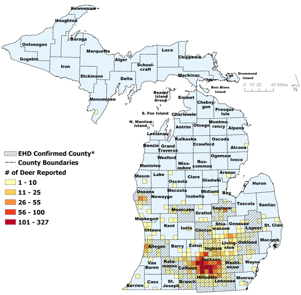
*Confirmed positives could be from lab results unrelated to reports made to the DNR using the online form.
Transmission
EHD is transmitted by Culicoides biting flies or midges, typically Culicoides variipennis. A deer must be bitten by a midge carrying the virus to become infected and the disease is not transmitted directly from one deer to another. Dead deer are not a significant source of virus since the virus does not survive long after the deer host dies.
In Michigan, outbreaks of EHD have occurred during late summer and early fall (August-October). Outbreaks typically end within two weeks of the onset of frost, since freezing temperatures kill the midge that transmits the virus.
Clinical Signs
Clinical signs of EHD are sudden and can be severe. Deer initially lose their appetite and fear of humans, grow progressively weak, become disoriented, may have excessive salivation, and develop a fever. With the high fever that they experience, deer seek out bodies of water to immerse themselves as they attempt to reduce their body temperature.
The hemorrhage that develops can result in bloody discharge from the nose or mouth, open mouth sores, or swelling of the eyelids, neck or head. A lack of oxygen in the blood can cause the tongue or inside of the mouth to become blue-tinged. Deer often develop shock, become recumbent, and die within 36 hours of the first clinical signs.
Although EHD infection can be fatal, some infected deer survive, including some that never show clinical signs. Deer that survive EHD infection may show hoof overgrowth with indentations or cracks in the wall that correspond to an interruption of hoof growth at the time of infection.
Pathology
The lesions of EHD are characterized by extensive hemorrhage as there is dysfunction of the body’s clotting mechanisms, damage to the blood vessel walls, and interference with blood circulation. Multiple organs are affected, particularly the heart, liver, spleen, kidney, lung, and intestinal tract.
Generalized tissue swelling from excess fluid accumulation is common including increased fluid around the heart.
Diagnosis
EHD is diagnosed through a combination of case history, characteristic signs and lesions, molecular testing, and/or or culture of the virus. EHD is highly likely when outbreaks occur in the late summer or early fall, deaths occur in or near water, and the dead deer have signs of hemorrhage or fluid accumulation.
The virus can be recovered from a variety of tissues, including blood, liver, spleen, kidney, lung, heart, and muscle. However, tissues need to be preserved for testing within 8-16 of the animal’s death.
Treatment and Control
There is no known effective treatment or control of EHD for wild populations of deer.
The DNR tracks reports of EHD through the Eyes in the Field online reporting system. Each year, the DNR will test EHD-suspect cases in a county until EHD is confirmed in that county in that calendar year. Once confirmed, DNR monitors reports of additional die-offs within that county but does not typically perform additional testing for that county that year.
Significance
Losses to EHD in a given area can be dramatic, and the sudden and pronounced loss of deer to EHD outbreaks can be upsetting.
Because of the high mortality rate, EHD can have a significant effect upon the deer population in a given area. However, studies conducted, including research performed in Michigan, have shown that outbreaks tend to be geographically localized and that the affected deer populations recover over the years following an outbreak. Recovery has occurred without restrictions on hunter harvest, but information is shared with the hunting community so that hunters and landowners can make informed decisions.
Although the EHD viruses can sometimes cause disease in cattle, sheep, and other domestic ruminants, livestock are much more commonly affected by the closely related Bluetongue Viruses than EHD viruses. Other domestic animals, such as dogs, cats, and horses, are not affected by EHD viruses, nor are people. Humans cannot contract EHD by any mode of transmission, including eating venison.
The DNR appreciates the public concern and willingness to share reports of deer disease and mortality.
For questions about wildlife diseases, please contact the Michigan DNR Wildlife Health Laboratory.
Photos
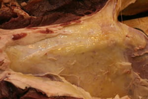

First figure: Edema (yellowish fluid) underneath the skin. Second figure: Hemorrhages on the organs, diaphragm, and musculature.